by Ravindra Warang
7 minutes
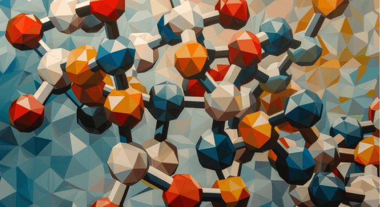
3D vs 2D Cell Culture: Which Model Is Right for Your Lab?
Explore how 3D cell culture is transforming drug discovery by offering more accurate, tissue-like models than traditional 2D methods.
A promising cancer therapy had cleared every preclinical hurdle. It killed tumor cells in culture, passed animal trials, and entered human testing with high hopes. But in Phase I trials, it failed—badly. Why?
The answer lay in the model used. The drug had been tested in flat, 2D cell culture—where cells spread unnaturally on plastic, isolated from real-world complexities. In patients, tumors aren’t flat; they’re dense, three-dimensional ecosystems known as the tumor microenvironment.
That trial became a turning point. Researchers realized that when your model doesn’t mimic the body, your results don’t translate. This realization paved the way for 3D cell culture—a shift from Petri dish biology to tissue-like realism. Techniques such as the hanging drop technique for spheroid formation have become essential in creating multicellular tumor spheroids (MCTS) that better represent cancerous tissues.
If you're a scientist, CRO, or lab planning your next experiment, this article will help you decide: Should you stick with 2D cell culture, or is it time to move into 3D? Discover applications of 3D cell culture in cancer research and explore how these advanced preclinical models can enhance drug discovery efforts.
Consider the limitations of traditional cytotoxicity assays for chemotherapy drugs that rely on 2D cell cultures. As seen with SW-480 cells using the CellTiter 96® Aqueous Non-Radioactive Cell Proliferation Assay Kit, results may not accurately reflect how tumors respond in vivo due to their simplified nature.
Embracing 3D cell culture techniques allows for a more accurate representation of cellular interactions and the effects of cytokines within the tumor microenvironment. By doing so, you’ll gain deeper insights into cell proliferation patterns and the intricate dynamics of cancer therapy response.
As you weigh your options between 3D vs 2D cell culture, remember that effective preclinical models are crucial for reliable outcomes in scientific research.
What Is 2D Cell Culture? Why Has It Been the Standard for So Long?
2D cell culture, often referred to as traditional cell culture methods, refers to the method of growing cells in a single layer on flat surfaces—flasks, Petri dishes, or multi-well plates. This approach has been fundamental in various scientific fields, including antibiotics research, vaccines development, and cancer biology studies.
Why it's been widely used:
- Inexpensive
- Easy to handle
- Standardized protocols
- Compatible with high-throughput screening (HTS)
For decades, 2D cultures have powered breakthroughs in antibiotics, vaccines, and cancer biology. However, they come with significant limitations.
Limitations of 2D cultures:
- Limited cell–cell interaction
- No spatial organization
- Drug efficacy overestimation
- Poor mimicry of human tissue response
These limitations of 2D cultures are increasingly relevant as researchers explore more advanced techniques like magnetic levitation in 3D cultures and precision medicine and personalized oncology.
2D models remain a workhorse in many labs, but their relevance is increasingly being questioned in modern, precision-driven R&D. As studies reveal differences in cytotoxicity responses between 2D and 3D cultured cells exposed to chemotherapy drugs, the need for more sophisticated models that accurately reflect drug metabolism and physiological conditions becomes clear.
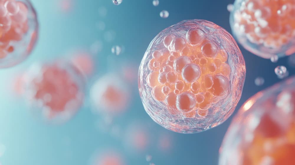
What Makes 3D Cell Culture Different?
3D cell culture is a technique that allows cells to grow in three dimensions, enabling them to expand in all directions and mimic their behavior in real tissues. This method is very different from traditional 2D cell cultures and offers several unique advantages.
Self-Assembly and ECM Interaction
These models self-assemble into structures such as spheroids and organoids, facilitating complex extracellular matrix (ECM) interaction.
Dynamic Engagement with Surrounding Cells
They dynamically engage with surrounding cells while creating natural gradients of oxygen (oxygen gradients), pH (pH gradients), and nutrients (nutrient gradients).
Realistic Environment for Disease Modeling
This realistic environment is crucial for accurate disease modeling architecture.
3D cultures provide:
- Better gene expression profiles in 3D cultures
- More accurate drug resistance behavior in 3D cultures
- Improved toxicological prediction in 3D cultures
Whether using scaffold-based methods, scaffold-free methods, organoid-driven cultures, or chip-integrated cultures, 3D cell culture simulates the in vivo environment far more effectively than 2D alternatives. Techniques like ultra-low attachment spheroid plates are essential for maintaining these complex structures during drug discovery processes.
By incorporating specific cell line culturing techniques and assessing outcomes through flow cytometry (FACSCalibur) or apoptosis analysis in 2D and 3D cultures, researchers can gain deeper insights into the differences between 2D and 3D culture dynamics.
3D vs 2D Cell Culture: A Comparison

This comparison of 2D and 3D cell culture highlights the significant differences in features such as growth patterns, cell–cell interactions, and drug sensitivity assessment in 2D and 3D environments. As researchers explore advanced techniques like Matrigel for creating more accurate extracellular matrices, the limitations of 2D cell culture models become increasingly apparent, especially in studies involving colon cancer cell lines in research.
When to Use 2D Culture — It’s Not Obsolete Yet
Despite its limitations, 2D cell culture still holds value in certain contexts:
✅ High-throughput screening (HTS) applications — early-stage compound elimination
✅ Cytotoxicity assays in 2D cultures — basic assessments of cell viability
✅ Genetic manipulations in 2D cultures (e.g., CRISPR knockouts in cell culture)
✅ Receptor-ligand interaction studies in 2D
Tip: Start with 2D to screen thousands of molecules quickly and cheaply using effective molecule screening techniques. Then validate the shortlisted ones using three-dimensional (3D) cell culture models.
Consider the complementary use of 2D and 3D models, especially when investigating the role of ECM in drug efficacy testing or understanding differences between 2D and 3D cell culture models for drug testing.
When to Use 3D Culture — Where Depth Delivers
Understanding when to use 3D cell culture is crucial for various research applications. 3D systems become essential when:
- The importance of tissue architecture matters (e.g., solid tumors, skin, liver)
- You're studying drug penetration in 3D models, hypoxia modeling in 3D, or conducting immune infiltration studies in 3D
- Gene expression fidelity in 3D is critical
- You’re modeling complex diseases like cancer or Alzheimer’s through complex disease modeling
- Personalized therapy testing is the goal (e.g., patient-derived organoids)
3D models excel in:
- Tumor microenvironment simulation
- Stem cell differentiation in 3D
- Toxicology and safety pharmacology in 3D
- Infectious disease modeling in 3D (e.g., SARS-CoV-2, Zika)
These applications highlight the unique advantages of 3D cultures over traditional methods, especially in fields like in vitro cancer models and cardiomyocyte research.
Real-World Use Cases — Who’s Using What and Why
Roche
Uses 3D tumor spheroids to model hypoxic tumor cores and test immunotherapies.
Emulate Inc.
Deploys liver-on-chip platforms for preclinical hepatotoxicity screening.
NIH + Harvard
Built brain organoids for Zika virus modeling to assess the impact of the Zika virus on fetal development.
MSKCC (Memorial Sloan Kettering)
Uses patient-derived organoids to match therapies to drug-resistant pancreatic cancer patients.
Insight: Most advanced labs now use a tiered approach in cell culture: 2D for screening → 3D for prediction → organoids for personalization. This approach addresses the limitations of 2D cell culture and enhances methods to assess drug efficacy using cell cultures.
Cost, Infrastructure, and Practical Considerations in Cell Culturing
Cost Comparison: 2D vs 3D Cell Culture

Infrastructure Requirements for 3D Culture
While 3D culture may demand more resources—such as polymeric hard material supports for cell culture—it often prevents costly failures later in the pipeline. This is particularly important when working with complex cell types like induced pluripotent stem cells or studying specific conditions such as colorectal adenocarcinoma cell lines.
Additionally, the use of vascular endothelial growth factor (VEGF) in 3D cultures can enhance the formation of realistic tissue models that better mimic in vivo environments. When considering practical considerations in cell culturing, it's essential to weigh the advantages of 3D cell culture against traditional methods like MSC spheroids vs 2D MSC cultures benefits.
As new technologies emerge—find out how new technologies like SeedEZ or magnetic levitation improve cell culturing—it's crucial to stay informed about the evolving landscape of cellular research and its applications in institutions like the Oncology Center Baghdad Teaching Hospital.
Future Outlook — Hybrid Workflows and AI Integration
The future is not 2D vs 3D — it’s 2D + 3D + AI.
- Labs are developing integrated platforms with both models, emphasizing hybrid workflows in cell culture.
- AI tools are enabling predictive analytics with AI based on 3D data, enhancing accuracy in gene expression analysis in cell cultures.
- Organoids are being tied to patient biobanks and organoids, allowing for personalized research applications.
- Regulatory bodies like FDA and EMA are including 3D data in submissions, highlighting the importance of regulatory acceptance of 3D data by FDA and EMA.
By 2028, most pharma R&D pipelines are expected to adopt multi-model workflows, combining:
- Flat models for speed, helping researchers understand differences between 2D and 3D cell culture methods
- 3D models for realism, particularly using materials like hydrogel and polymeric hard scaffolds
- Organoids for personalization, especially in studies involving mesenchymal stem cells (MSCs) and growth factors like platelet-derived growth factor (PDGF)
These advancements will not only streamline processes but also reduce reliance on animal models in research, as seen with Caco-2 studies that assess drug efficacy using compounds like Doxorubicin (Doxo).
Conclusion: Flat Biology vs Real Biology — Choose Your Lens Wisely
Think of 2D models as sketches — quick, cheap, and informative. But they miss depth in biological modeling, detail, and nuance. 3D models, on the other hand, offer blueprints — detailed, realistic, and often predictive of real-life outcomes, especially in cell culture.
For a lab, the strategic choice between 2D and 3D models isn’t binary — it’s strategic. Know what you’re trying to uncover. Match your model to your research question. For instance, when exploring cancer research or drug development using colorectal cancer cell lines like HCT-116, consider how techniques such as organoid culture and disease modeling can provide insights that flat models might miss. Be ready to evolve with advancements like magnetic levitation or hanging drop microplates.
Because in modern biology, depth matters — not just in science, but in the systems we use to explore it.
FAQs (Frequently Asked Questions)
Q1: What is the difference between 2D and 3D cell culture?
The difference between 2D and 3D cell culture lies in their structural environments: 2D cell culture grows cells on flat surfaces, while 3D culture allows cells to grow in all directions, mimicking tissue-like behavior. This spatial growth is crucial for studying the impact of extracellular matrix (ECM) components on cellular behavior and drug response.
Q2: Which is better — 2D or 3D cell culture?
It depends on the research goals. 2D cell culture is ideal for basic screening due to its simplicity and cost-effectiveness—these are some of the advantages of 2D cell culture. In contrast, 3D cell cultures offer better physiological relevance and predictive power for drug discovery, making them essential for studies in oncology and regenerative medicine.
Q3: Is 3D cell culture more expensive than 2D?
Yes, 3D cell culture methods are generally more expensive than traditional 2D approaches. Cost comparison of cell culture methods reveals that 3D systems require specialized media, equipment like spheroid microplates, and handling expertise. However, they often provide more accurate results, particularly in complex biological assays.
Q4: What industries are adopting 3D cell culture?
Industries adopting 3D cell culture include pharma, biotech, contract research organizations (CROs), and academic labs. These sectors utilize advanced models for research areas like neuroscience, toxicology, and drug development. As the field evolves, the demand for innovative techniques such as scaffold-free 3D culture methods continues to rise.
Q5: What are some challenges faced when transitioning from 2D to 3D cell culture models?
Challenges include higher costs, increased complexity in experimental design, and the need for specialized training to handle techniques effectively. Researchers must also consider factors like nutrient diffusion limitations and variations in cellular heterogeneity within different types of 3D cultures.