3D Cell Culture Explained: Types, Applications, and Emerging Trends in Pharma
Discover how 3D cell culture is transforming pharma with organoids, bioprinting, and human-relevant drug testing models.

Key Takeaways
- 3D cell culture bridges the gap between in vitro experiments and in vivo biology, offering more accurate, ethical, and human-relevant models.
- It encompasses a spectrum of technologies — from simple spheroids to organoids, microfluidic organ-on-chips, and 3D bioprinted tissues.
- Pharma, biotech, CROs, and academia are rapidly adopting 3D models for drug discovery, toxicology, neuroscience, immuno-oncology, and personalized medicine.
- The global market is growing at 18–22% CAGR, driven by demand for better translational models and the move away from animal testing.
- Challenges remain around scalability, standardization, and cost, but advances in AI, automation, and regulatory support are accelerating adoption.
- 3D cell culture is no longer experimental — it is foundational to the future of precision medicine and next-generation therapy development.
Introduction
"In 2003, a team of cancer researchers in Boston noticed something odd. The same breast cancer cells they had studied for years in petri dishes began behaving completely differently when grown in a gel-like matrix. They clustered, formed tight colonies, and even responded to drugs they once ignored. What changed? The cells weren't growing flat anymore. They were growing in 3D."
This seemingly simple shift — from flat to dimensional — marked a turning point in how scientists began to understand biology outside the body. The real world, after all, isn't flat. Tissues curve, cells talk to each other in every direction, and oxygen doesn't flow evenly across every surface. Yet, for decades, scientists relied on 2D cultures to test drugs, model diseases, and predict human outcomes.
That's where 3D cell culture enters the frame.
Unlike traditional methods, 3D cell culture allows cells to grow, interact, and evolve in three dimensions — mimicking the microenvironments of real tissues. Today, it's revolutionizing everything from cancer drug screening to brain organoid development, and pushing the boundaries of what's possible in personalized medicine, regenerative therapy, and next-gen pharma R&D.
In this guide, we'll uncover:
- The science behind 3D cell culture (and why it matters),
- The types of systems and models in use today,
- Real-world case studies and research breakthroughs,
- And how 3D cultures are shaping the future of pharma and biotech.
Backed by expert insights, peer-reviewed data, and stories from the lab bench, this isn't just another technical explainer. It's your deep dive into one of the most impactful innovations in modern biology.
I. What Is 3D Cell Culture?
When we talk about scientific breakthroughs, most people think of gene editing, AI in drug design, or CRISPR. But often, it's the quiet revolutions — the ones that change how we study biology — that create the biggest shifts.
3D cell culture is one such shift.
For decades, researchers relied on flat, plastic surfaces to study cells — petri dishes, flasks, multi-well plates. These 2D models helped launch modern biology. But there's one problem: the human body isn't flat.
Cells in our body grow in a three-dimensional matrix — they stretch, pull, talk to neighbors, respond to forces, and live within gradients of oxygen, nutrients, and waste. When we study them on a flat surface, we're essentially asking them to behave unnaturally.
3D cell culture corrects this disconnect.
It gives cells the freedom to behave like they do inside tissues — not on lab plastic.
In its simplest form, 3D cell culture refers to techniques that allow cells to grow and interact in all directions, forming clusters, networks, or even miniature organ-like structures. These setups are designed to replicate the physical and biochemical cues of the extracellular matrix (ECM) — the invisible framework that holds our tissues together and guides cell behavior.
Why It Matters: From Petri Dishes to Precision Biology
The moment you switch to 3D, something remarkable happens:
- Cancer cells begin forming solid tumor spheroids, revealing how drug penetration varies by layer.
- Liver cells start producing albumin and cytochrome enzymes — just like they do in the body.
- Neurons extend into networks that fire signals — essential for neurodegenerative disease modeling.
In short, 3D cultures don't just look different — they behave differently, and those behaviors often mirror what happens inside the human body far more accurately than 2D.
For pharmaceutical R&D teams, this means:
- More predictive drug screening
- Lower chances of clinical trial failure
- Better models for toxicology, metabolism, and disease progression
For scientists, it means a richer, more realistic platform to explore biology's complexity.
And for the industry at large, it signals a pivot toward ethically sound, mechanistically relevant, and human-centric experimentation.
Next, we'll explore the different ways this is achieved — from scaffold-based matrices to self-assembling organoids.
II. Types of 3D Cell Culture Models
The power of 3D cell culture lies in its flexibility. It's not a one-size-fits-all method — it's a toolbox of strategies, each designed to mimic different aspects of living tissues.
Depending on your scientific question — whether it's understanding tumor invasion, testing hepatotoxicity, or modeling neural networks — the 3D system you choose can look vastly different.
Let's explore the major types of 3D cell culture models that are transforming lab benches across the globe.

1. Scaffold-Based Systems
Think of these as artificial extracellular matrices — materials that physically support cells, allowing them to grow, migrate, and differentiate like they would in tissues.
Scaffolds can be:
- Natural polymers like collagen, gelatin, alginate, or Matrigel™ — biologically active and rich in cell-binding sites.
- Synthetic polymers like PEG, PLA, or PLGA — tunable for stiffness, porosity, and biodegradability.
Cells are seeded into these 3D networks, where they form complex arrangements — from chondrocytes in cartilage models to fibroblasts in wound healing assays.
"Scaffold-based models are ideal when mechanical cues or tissue-like architecture is critical to your research."
2. Scaffold-Free Systems
Sometimes, you don't need a structure — the cells build it themselves.
In scaffold-free systems, cells aggregate naturally into spheroids or microtissues, relying on their own adhesion and signaling cues. These are commonly used in:
- Cancer biology (tumor spheroids)
- Stem cell differentiation
- Immune cell interaction models
Popular methods include:
- Hanging drop plates
- Ultra-low attachment (ULA) plates
- Rotating bioreactors and spinner flasks
These models allow tight cell–cell interactions, nutrient gradients, and hypoxic cores — making them ideal for studying drug penetration and resistance mechanisms in solid tumors.
3. Organoids: The Mini Organs of the Lab
If spheroids are simple 3D structures, organoids are biological symphonies.
Derived from stem cells or patient biopsies, organoids are self-organizing, multicellular structures that mimic the architecture and function of real organs — think mini-guts that absorb nutrients, liver organoids that detoxify drugs, or brain organoids with firing neurons.
They are a breakthrough in:
- Personalized medicine
- Developmental biology
- Rare disease modeling
- Infectious disease research (e.g., COVID-19 studies using lung organoids)
"Organoids are where 3D culture meets regenerative biology — highly complex, ethically powerful, and deeply human."
4. Organ-on-Chip & Bioprinting Technologies
At the cutting edge are microengineered systems that go beyond passive culture — they simulate fluid flow, mechanical forces, and inter-organ communication.
- Organ-on-chip platforms use microfluidic devices lined with human cells — allowing real-time observation of tissue responses.
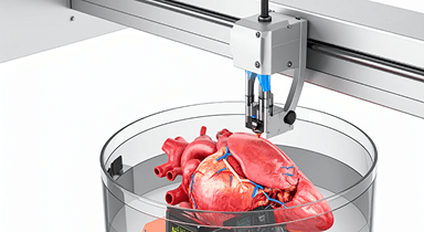
- 3D bioprinting enables scientists to print tissues layer-by-layer using bioinks containing cells and ECM components.
These tools are being adopted in toxicology, pharmacokinetics, and multi-organ interactions, including human-on-chip models that represent entire biological systems.
Choosing the Right Model: It Depends on the Question

Each model comes with its strengths and trade-offs. But together, they offer an unprecedented opportunity to explore biology in ways that were once only possible inside a living organism.
Up next, we'll explore how these models are applied across pharma, biotech, and academic research — and why they're changing how we approach everything from cancer to cardiotoxicity.
How Organoids Are Shaping the Future of Personalized Medicine
Read moreIII. Applications of 3D Cell Culture in Pharma and Life Sciences
"In drug development, 90% of drugs that look promising in preclinical studies fail in human trials."
One major reason? Traditional 2D cultures fail to replicate how real tissues respond.
3D cell culture is changing that.
By mimicking the structural and functional nuances of living tissues, 3D systems are offering more predictive, ethical, and cost-effective models. Whether you're modeling a metastatic tumor, testing liver toxicity, or simulating immune responses — 3D culture is no longer a futuristic concept. It's happening now.
Let's look at how this technology is being applied across domains.
1. Drug Discovery and Preclinical Testing
The pharmaceutical industry has long struggled with the translational gap — the mismatch between how a drug performs in lab models versus in patients.
3D models reduce that gap by offering:
- Realistic tissue penetration studies (e.g., anti-tumor drugs)
- In vitro drug metabolism and clearance testing using liver organoids
- Dose-response curve accuracy far superior to 2D
Companies are now integrating 3D liver spheroids, cardiac microtissues, and lung-on-chip systems into early-stage screening — helping flag failures before they reach costly human trials.
Example: Roche and AstraZeneca have both published studies showing improved predictability using 3D tumor models in oncology pipelines.
2. Cancer Research & Tumor Microenvironment Modeling
Cancer isn't just about rogue cells — it's about how those cells interact with blood vessels, immune cells, and extracellular matrix.
3D cultures help simulate this complexity:
- Tumor spheroids mimic hypoxic cores and drug gradients
- Co-culture systems replicate immune–tumor interactions
- Patient-derived organoids (PDOs) predict personalized therapy outcomes
"In 2D, cancer cells die when they're supposed to. In 3D, they fight back — like in real tumors."
This realism is vital for immunotherapy testing, checkpoint inhibitor evaluation, and resistance mechanism research.
3. Toxicology and Safety Pharmacology
Regulatory bodies like the FDA are increasingly advocating for non-animal, human-relevant testing.
3D systems shine here:
- Hepatotoxicity: Liver spheroids show dose-dependent toxicity and enzyme induction.
- Cardiotoxicity: Cardiac tissues beat in vitro — allowing real-time arrhythmia detection.
- Nephrotoxicity: Kidney organoids filter and reabsorb fluids, helping assess renal drug impact.
Many pharma companies now maintain 3D safety panels alongside traditional animal models to support IND submissions.
Using 3D Cell Culture for Toxicity Testing: A Step-by-Step Framework
Read more4. Neuroscience and Brain Research
3D neural cultures and brain organoids have become essential in:
- Modeling neurodevelopmental disorders like autism and microcephaly
- Studying neurodegeneration (e.g., Alzheimer's plaques in 3D cultures)
- Investigating viral infections (e.g., Zika virus damaging neural progenitors)
These models even exhibit electrical activity and synaptic communication, enabling functional testing that's impossible in 2D.
5. Stem Cell Research and Regenerative Medicine
From intestinal crypts to retinal patches, stem-cell-derived organoids are being used to:
- Study organ development and genetic diseases
- Test gene therapies in patient-specific models
- Produce implantable tissues for corneal, liver, and cardiac repair
Bioengineered 3D tissues could soon become mainstream in clinical transplantation pipelines.
6. Infectious Disease and Virology
The COVID-19 pandemic highlighted the power of 3D systems:
- Lung organoids were used to model SARS-CoV-2 entry and immune response
- Intestinal organoids helped study GI symptoms and viral replication
- Viral efficacy testing was performed without relying solely on animal models
This opens doors to faster, safer infectious disease modeling — especially for pathogens affecting human-specific receptors.
7. Personalized Medicine & Biobanking
Imagine testing therapies on a miniature replica of a patient's tumor or liver before giving them the real drug.
That's what 3D patient-derived models enable:
- Match the right treatment to the right patient
- Identify resistance patterns early
- Build living biobanks of tumors for large-scale testing
Institutions like the Hubrecht Organoid Technology (HUB) are already leading global efforts to integrate organoids into personalized cancer care.
3D cell culture isn't just a lab innovation anymore.
It's a clinical ally, a regulatory companion, and a window into human biology like we've never had before.
IV. 3D vs 2D Cell Culture – A Comparative View
For decades, 2D cell cultures were the gold standard. They gave us penicillin breakthroughs, early vaccine models, and even paved the way for stem cell research.
But biology, like innovation, isn't flat.
As the complexity of questions in drug development and disease modeling deepens, scientists are turning to models that closely replicate in vivo conditions. This is where 3D cell cultures rise to the occasion — offering not just better science, but more human science.
To truly understand the leap, here's a direct comparison:
3D vs 2D Cell Culture Comparison Table

Why This Comparison Matters
Many R&D teams still start with 2D for cost efficiency and simplicity. But as they progress to later stages — where human relevance becomes critical — 3D systems offer the next layer of validation.
In fact, leading pharmaceutical companies are building hybrid pipelines:
- 2D for initial compound screening
- 3D for predictive toxicology and validation
- Organoids for patient-matched testing
"Think of 2D as the sketch. 3D is the blueprint."
Both are useful. But when precision, realism, and patient safety matter — 3D changes the game.
3D vs 2D Cell Culture: Which Model Is Right for Your Lab?
Read moreNext, let's ground this comparison in real-world stories. Who's using these models today? What breakthroughs are already happening?
V. Case Studies and Real-World Usage of 3D Cell Culture
In the world of science, credibility comes not just from theory — but from who's using it, how, and what it changes.
Over the past decade, 3D cell culture has moved from academic journals into the core workflows of pharmaceutical R&D labs, CROs, and biotech startups. Below are selected examples that highlight how this technology is shaping results, reducing risk, and enabling faster, safer decisions.
Case Study 1: Roche — Advancing Tumor Response Modeling
Context: Roche's oncology division was struggling with low correlation between in vitro drug data and actual tumor response in patients.
Solution: The company adopted 3D tumor spheroids made from patient-derived cancer cells, especially for breast and lung cancer compounds.
Outcome:
- Improved accuracy in predicting drug penetration and resistance
- Reduced false positives in early drug screening
- Enabled faster go/no-go decisions in the preclinical stage
Lesson: 3D spheroids helped replicate the hypoxic tumor core, making drug efficacy testing more reliable.
Case Study 2: Emulate Inc. – Organ-on-Chip for Liver Toxicity
Context: Many drugs fail in late-stage trials due to hepatotoxicity undetected in animal models or 2D cultures.
Solution: Emulate partnered with major pharma companies to deploy liver-on-chip models. These microfluidic systems used human hepatocytes with fluidic flow to mimic blood circulation.
Outcome:
- Detected drug-induced liver injury (DILI) missed by 2D models
- Used in regulatory submissions as supplementary evidence
- Reduced reliance on animal studies
Insight: The chip even predicted toxic responses 7 days before visible signs appeared in vivo.
Case Study 3: HUB Organoids – Personalized CRC Treatment
Context: A colorectal cancer (CRC) patient was resistant to standard chemotherapy. Time was running out.
Solution: A biopsy was used to grow a patient-derived organoid (PDO) within 2 weeks. Multiple drug combinations were tested on this mini-tumor in the lab.
Outcome:
- One combination showed a strong apoptotic response in the organoid
- Clinician administered that same combo to the patient
- Patient responded with partial tumor shrinkage and improved prognosis
Takeaway: This was personalized medicine in action, using 3D culture to inform real-time clinical decisions.
Case Study 4: NIH & Harvard – Brain Organoids for Zika Research
Context: The Zika virus epidemic raised concerns about microcephaly in infants. Animal models failed to explain the human-specific mechanism.
Solution: Researchers used 3D brain organoids derived from human iPSCs to simulate fetal brain tissue.
Outcome:
- Discovered that Zika specifically targets neural progenitor cells
- Validated the link between virus exposure and impaired brain development
- Informed public health strategy and maternal guidelines
Lesson: 3D organoids provided a human-specific window into neurodevelopmental damage — without needing human fetal tissue.
Case Study 5: Takeda – 3D Culture for Immuno-Oncology
Context: Takeda's immuno-oncology team needed to evaluate how T cells infiltrate solid tumors — something 2D co-cultures couldn't model well.
Solution: Adopted immune-enhanced tumor spheroids, embedding tumor and immune cells in hydrogel matrices.
Outcome:
- Quantified immune cell infiltration and cytokine release
- Prioritized compounds for checkpoint blockade therapy
- Improved translational alignment with human tumor biology
Insight: These assays reduced the need for murine syngeneic models, accelerating early-stage decisions.
What These Cases Show
- Pharma giants are integrating 3D culture into core pipelines — not just experiments
- CROs are offering 3D assays as a competitive differentiator
- Clinicians are increasingly interested in lab-to-clinic personalization
"The gap between what works in a dish and what works in a human is closing — thanks to 3D."
Inside a 3D Cell Culture Lab: Equipment, Costs & Setup Guide
Read moreIn the next section, we'll explore how this momentum is reflected in global trends — funding, demand, innovation, and what's coming next.
VI. Market Trends and Future Outlook of 3D Cell Culture
"What began as a research curiosity is now a billion-dollar industry redefining how we study, treat, and personalize healthcare."
A Market in Motion
The global 3D cell culture market is on a steep upward trajectory. Once confined to academic labs, it's now a strategic investment area for pharma companies, CROs, biotech firms, and regulatory science organizations.
Global Market Snapshot (2024–2025)

Key Drivers:
- Rising failures in late-stage drug trials
- Demand for ethical alternatives to animal testing
- Explosion in personalized and precision medicine
- Adoption of AI-integrated organoid platforms
- Regulatory support for human-relevant data models
Who's Investing in 3D Cell Culture?
Pharma & Biotech
Major players like Roche, Novartis, Pfizer, AstraZeneca, and Takeda are either building internal 3D culture programs or partnering with platform providers.
CROs & Platforms
Companies like:
- InSphero (Switzerland): Advanced tumor and liver spheroid models
- Emulate Inc. (USA): Organ-on-chip systems for pharma safety
- MIMETAS (Netherlands): High-throughput microfluidic organ systems
- Hubrecht Organoid Technology (HUB): Patient-derived organoid biobanking
They're offering 3D culture as a service model — much like sequencing was in the 2010s.
Investors
Venture capital is pouring in:
- Emulate Inc. has raised over $150M+
- Cellink/BICO Group (3D bioprinting leader) is expanding aggressively
- Japanese and EU biotech investors are focusing on organoid drug screening startups
Trends That Are Shaping the Future
1. Patient-Derived Organoid Biobanks
Hospitals and CROs are building biobanks of tumor, liver, gut, and brain organoids — allowing:
- Population-scale drug testing
- Rare disease modeling
- AI-powered phenotype mapping
HUB, Candiolo Cancer Institute, and MD Anderson are leading this wave.
2. AI + 3D Cell Culture Integration
The convergence of AI-based imaging, machine learning, and 3D culture data is enabling:
- Automated organoid classification
- Predictive modeling of drug efficacy
- Faster compound selection through phenotype clustering
Startups like Curi Bio and Tara Biosystems are pioneering AI-integrated platforms.
3. Regulatory Recognition is Growing
- The FDA Modernization Act 2.0 (2023) encourages non-animal methods in drug development.
- EMA and OECD are expanding guidelines to include 3D-based in vitro methods for toxicity, DMPK, and mechanistic studies.
Implication: 3D data is now seen as complementary to animal studies, and in some areas, preferable.
4. Organ-on-Chip Ecosystems for Multiorgan Simulation
Imagine testing drug absorption in the gut, metabolism in the liver, and toxicity in the heart — all connected via microfluidics.
These "Human-on-Chip" systems are:
- Being tested by NASA for space biology
- Piloted by the NIH's Tissue Chip for Drug Screening program
- Integrated into pharma workflows for systems biology modeling
5. 3D Bioprinting for Transplantable Tissues
Beyond research, 3D cell culture is paving the path for engineered tissues:
- Cardiac patches for ischemic patients
- Corneal layers for eye surgery
- Bone scaffolds for maxillofacial repair
3D Bioprinting in Regenerative Medicine: Hype vs Reality
Read moreThough not yet in clinical practice at scale, these are at advanced preclinical stages.
The Decade Ahead: What To Expect

"3D culture won't just be a tool for research — it will be a clinical compass guiding real-world treatment decisions."
VII. Challenges and Limitations of 3D Cell Culture
"Every leap in science comes with friction — not because it's wrong, but because it's new, nuanced, and disruptive."
While 3D cell culture offers incredible promise, it's not a silver bullet. Labs, pharmaceutical companies, and CROs that adopt it face practical challenges that still need solutions. Here's an honest look at the current roadblocks in adoption and scalability.
1. Scalability and Reproducibility
The Challenge:
Most 3D culture methods — especially organoids and bioprinted tissues — are labor-intensive and manually handled. This leads to:
- Batch-to-batch variability
- Irregular size, shape, or maturation of organoids
- High variability in drug response
Why it matters: Pharma R&D and regulatory submissions depend on standardization and repeatability. If two labs produce different results using the same protocol, confidence erodes.
Emerging Solutions:
- Automated bioreactors (e.g., CelVivo, CERO)
- AI-based organoid tracking and classification
- High-content screening-compatible 3D plates (Greiner Bio-One, Corning)
2. Cost and Infrastructure Demands
The Challenge:
3D cultures can be 2–5 times more expensive than 2D setups. They require:
- Specialized media, matrices (e.g., Matrigel™)
- Microfluidic devices or 3D bioprinters
- Skilled personnel for handling and interpretation
For many mid-size CROs or academic labs, the capital and training investment remains a hurdle.
Workarounds:
- Shared platforms and consortia (e.g., HUB, EBiSC)
- 3D assay outsourcing to service providers
- Open-source bioprinting platforms (like CELLINK Bio X community)
3. Data Complexity and Interpretation
The Challenge:
3D cultures generate nonlinear, high-dimensional data:
- Z-stacks of imaging data
- Real-time functional outputs (beating cardiomyocytes, synapse activity)
- Spatial heterogeneity within spheroids or organoids
This data is rich but hard to interpret using standard 2D analysis tools.
Solutions on the Horizon:
- AI-assisted phenotypic analysis tools (e.g., DeepCell, CytoReason)
- 3D-specific statistical models and machine learning pipelines
- Integration with transcriptomics and proteomics platforms
4. Lack of Universal Protocols
The Challenge:
Every lab tweaks protocols based on cell type, matrix, or medium — leading to:
- No gold standard for reproducibility
- Limited peer-to-peer comparability
- Regulatory skepticism
Why it matters: Without harmonized SOPs, 3D data risks being dismissed as "scientific black boxes" by regulators and pharma QA teams.
Response:
- Initiatives by the OECD and FDA to establish 3D validation guidelines
- Academic–industry consortia pushing for reference SOPs (e.g., EU ORCHID Project)
5. Not Always the Right Model
The Challenge:
3D systems are more realistic, but not always necessary. For:
- High-throughput, early screening of thousands of compounds
- Basic cytotoxicity checks or receptor-ligand binding studies
2D models remain faster, cheaper, and good enough.
"The key is knowing when to use 3D — not just using it for the sake of innovation."
Many labs adopt a tiered approach:
- Use 2D for volume screening
- Use 3D for predictive modeling and clinical-stage validation
Final Thought on Challenges
Innovation is never perfect at birth. Just as PCR and next-gen sequencing faced initial skepticism, 3D cell culture is undergoing its own phase of scientific maturation.
The community — from academic labs to regulators and startups — is already responding with:
- Smarter instruments
- Interoperable datasets
- Collaborative frameworks
What was once an "elite" research method is on its way to becoming standard practice — and the hurdles are part of that journey.
Next, let's showcase who's driving this evolution — from companies and startups to academic powerhouses.
VIII. Leading Companies, Institutions, and Innovators in 3D Cell Culture
"Innovation doesn't happen in isolation. It takes an ecosystem — and 3D cell culture now has one."
From bioprinting startups and organoid pioneers to academic labs and pharma alliances, 3D cell culture is no longer niche. It's a fast-growing field being shaped by cross-sector collaboration, venture-backed platforms, and scientific leaders who are redefining how we study biology.
Here's a closer look at the players driving the 3D cell culture revolution — and what makes them stand out.
Top Companies in 3D Cell Culture

1. InSphero AG (Switzerland)
- Focus: 3D microtissues for liver, pancreatic, and tumor models
- Why they matter: Their GravityTRAP™ and Akura™ Plates enable scalable 3D assays for pharma use.
- Clients: Used by Novartis, Merck, and several FDA partners
2. Emulate Inc. (USA)
- Focus: Organ-on-chip platforms
- Why they matter: Their liver and intestine chips are already used in regulatory toxicology and pharma R&D
- Backed by: DARPA, NIH, and venture funds; spun out of Harvard Wyss Institute
3. MIMETAS (Netherlands)
- Focus: OrganoPlate® – a microfluidic 3D culture platform
- Why they matter: Enables high-throughput organ-on-a-chip testing without pumps
- Used for: Drug screening, ADME/Tox, and disease modeling
4. Corning Life Sciences
- Focus: ECM products (like Matrigel™), 3D culture plates
- Why they matter: A legacy labware giant that supplies the scaffolds and substrates for many global 3D projects
5. Cellink/BICO (Sweden)
- Focus: 3D bioprinting and tissue engineering
- Why they matter: Their Bio X™ printer is one of the most widely used desktop bioprinters in the world
6. Hubrecht Organoid Technology (HUB)
- Focus: Patient-derived organoid biobanks
- Why they matter: One of the first groups to commercialize PDOs for personalized oncology and pharma validation
- Spin-out from: Hubrecht Institute, Netherlands
Academic Institutions Leading the Field

Global Collaboration and Consortia
Tissue Chip for Drug Screening (USA)
NIH-led program supporting the development of multiple organ-on-chip systems for pharma pipelines
ORCHID Consortium (EU)
Pan-European project to standardize organ-on-chip validation and regulatory readiness
EpiOrganoid Alliance (Industry + Academia)
Global alliance focused on epithelial tissue modeling for cancer and respiratory diseases
Japan Organoid Consortium
Government-backed push to scale bioprinted tissues for clinical and cosmetic testing
Innovators to Watch (Startups & Labs)

Why This Ecosystem Matters
3D cell culture is not just a product or technique — it's an ecosystem of enablers:
- Labware manufacturers making specialized scaffolds
- Software companies building AI analysis tools
- Biobanks storing patient-specific mini-organs
- Academic spinouts turning discoveries into platforms
The real innovation lies in how these players collaborate — where pharma meets deep tech, and where biology becomes programmable.
IX. Frequently Asked Questions (FAQs)
What is 3D cell culture, and how is it different from 2D cell culture?
3D cell culture allows cells to grow in all directions, simulating the structure and function of tissues inside the body. In contrast, 2D cell culture grows cells on flat surfaces, often leading to unnatural morphology and limited cell–cell interaction.
Why is 3D cell culture important in drug discovery?
3D models mimic real tissue environments, which means they offer more accurate predictions for drug efficacy, toxicity, and resistance. This leads to better preclinical screening, reduced reliance on animal models, and fewer late-stage failures.
What types of 3D cell culture systems exist?
There are four major types:
- Scaffold-based systems (e.g., collagen, Matrigel™)
- Scaffold-free systems (e.g., spheroids, hanging drops)
- Organoids (self-organizing mini-organs from stem cells)
- Organ-on-chip / Bioprinting (microfluidic and engineered tissue platforms)
Each has specific use cases in cancer, neurology, toxicology, and regenerative medicine.
Can 3D cell culture replace animal testing?
Not entirely — yet. But it's increasingly used as a complementary or alternative model, especially in early drug development, toxicity studies, and personalized medicine. Global regulatory bodies like the FDA and EMA are recognizing its value.
What are organoids, and how are they made?
Organoids are miniature, 3D structures that mimic the architecture and function of real organs. They are typically derived from pluripotent stem cells or patient biopsies, and self-organize when placed in the right culture medium and ECM-like materials.
Are 3D cell culture models expensive?
Yes, they are generally more costly than 2D models — both in terms of materials and labor. However, they offer better predictive accuracy, reducing downstream costs associated with failed drugs or redundant animal testing.
How long does it take to grow organoids?
Depending on the cell type and complexity, organoids can form in 7–21 days. Brain or cardiac organoids may take longer to mature and require specific culture conditions.
Is 3D cell culture used clinically today?
What are the limitations of 3D cell culture?
- Scalability and reproducibility challenges
- High cost and specialized equipment
- Lack of standardized protocols
- Complex data interpretation
Despite these, the field is rapidly advancing with AI, automation, and cross-disciplinary innovation.
Which companies are leading in 3D cell culture innovation?
Some of the top names include:
InSphero, MIMETAS, Emulate Inc., Corning Life Sciences, Cellink (BICO), and Hubrecht Organoid Technology (HUB).
These companies provide platforms, biobanks, and devices that support global adoption of 3D cell culture systems.
X. Conclusion: From Flat Biology to Full-Scale Impact
"The future of life sciences won't be built on plastic plates — it will be built in dimensions that reflect the complexity of life itself."
3D cell culture is not just a new technique. It's a shift in perspective — one that brings us closer to human biology, human outcomes, and ultimately, human healing.
Whether you're a researcher looking to model disease more effectively, a pharma innovator trying to reduce clinical trial attrition, or a policy-maker shaping the next generation of regulatory frameworks — 3D cell culture is the bridge between biology and reality.
And that bridge is already being crossed.
Want More?
Looking to feature your 3D culture innovation or platform in our upcoming biotech spotlight? Reach out.
Need help writing for your life sciences brand? We specialize in science storytelling that connects.
References:
- MarketsandMarkets
- BCC Research
- Technavio
- FDA Modernization Act 2.0 Overview
- PubMed Article on FDA Modernization Act 2.0
- Emulate Inc. Liver-Chip
- Emulate Inc. Study Publication
- HUB Organoids
- InSphero 3D InSight™ Human Liver Microtissues
- Stanford's Vascularized Heart Organoids
- Hubrecht Institute Organoid Group